Saturday, November 1, 2014

Friday, 31st october 2014, "Algae And Fungi"

Since Dr Wan couldn't find the time to teach us Algae and Fungi on Monday nor Tuesday. Today we had a 4 hours long class. It was fun actually. I must admit that I'm starting to love Microbiology more. It's a good thing! Not that I hate it before but the fondness towards the subject is subtle before this. In my opinion, it's important to love what you're doing/learning. Same goes for your career in the future, just saying. x'D (Self reflection section haha) 

Since last week I have hmm somehow talked about algae. So for this week's entry, I will do the "New Things That I Learned Today" section! ^^ 

New Thing That I Learned Today

ALGAE
  • Algae is the first producer of the marine food chain.

  • Chlorophyta, Rhodophyta and Phaephyta are in the kingdom of Plantae.

  • Microbes that lives on neustonic region (at water-atmosphere interface) are autotrophic or photoautotrophic microbes because they need sun to produce food for themselves. 

  • Microbes that lives on benthic region (at the bottom of water) are anaerobes because at the bottom of water, there are low concentration of oxygen. 

  • Endosymbionts : Any organisms that lives in the cell of other organisms and interacting with each other by mutualism. 

  • Algal bloom : Although we often say things like "Wow! The lake is green! So pretty!" The green lakes are actually cause by algal bloom. It is toxic and can kill fish. (I will never think about green lakes the same way anymore...) (・⊝・)

  • Secondary product : Product that is produced by metabolism of algae or any other microbes. (Such as alginic acid, carrageenan etc) 

  • Algal bloom occurs when the lake is high in nutrition that will stimulate excessive plant growth (for example : algae). Also known as "eutrophication". 

  • The summarise consequences of algal bloom is that the excessive growth of algae will prevent the sunlight to penetrate the algae and into the water. This will lower the level of oxygen inside the lake. This will also highly stimulate the growth of anaerobes. The anaerobes will release toxic gases due to chemical changes and lastly kill marine organisms. 

  • Dinoflagellates produce brevetoxins which can cause red tides

  • The most studied mold is "slime molds". 

FUNGI
  • An example on the uses of fungi is it can be used as antibiotic. For example, cortisone which is a type of steroid that can treat a variety of skin condition such as rash, allergies, prevent swelling or redness due to insect bites and prevent inflammation. 

  • Anamorphic fungi produce asexual spores ONLY. 

  • Ascomycota, a type of fungi, reproduce asexually by conidia (asexually produced spores) called conidiospores

  • Stachybotrys cause "sick building syndrome" where usually we can see it on the ceiling or the walls.   
  • The fungus that looks like an ear here is called Auricularia auricula-judae

  • Microspodia is an obligate intracellular fungal parasites which means that they need host to survive. 

  • Mycorrhizae helps to absorb nutrient. The hyphae of fungi will form a sheath around the roots of plant, using its mycelium. The fungal-root coating is called "mycorrhizae". They can absorb water and mineral deeper into the soil that the plant's root cannot reach. 

  • Lichens can act as pollution indicators because they are sensitive to air pollution or environmental toxins. Thus, when there is lichens, it is free from pollutant. 

  • Algae is a photobiont while fungi is a mycobiont.  

  • Facultative : with or without oxygen, can still survive. 

  • Dimorphic fungi can form two types of growth which is mold-like or yeast-like. And it depends on the temperature and carbon dioxide


Monday & Tuesday, 27th & 28 October 2014, "Of Posters, Videos And Exhibition"

Monday, 27th October 2014

Since tomorrow we are having the long-awaited exhibition to present our long-awaited posters and videos. A replacement class was held today. But we didn't get to learn anything today because, since we will be viewing our videos tomorrow using the Aurasma application that we have installed on our devices, Dr Wan wanted to make sure everybody's Aurasma is working well and all the videos can be played. Andddd it actually took the whole two hours to settle this. Dr Wan was worried too as to how will we cover the subjects in order to finish it before the final examination. 

Tuesday, 28th October 2014, "Day Of The Exhibition!!" 

Our group's morning wasn't having a good start because our video suddenly couldn't be played on Aurasma. Hence, we had to put the video in Aurasma Studio all over again. And the internet at the faculty was not helping us at all x'D It took about 1 hour and a half for our video to be uploaded into the web. Thank you to some of my classmates that have helped us!! For 'lending' us their internet huddle and also Chew!! Helping us solve the problem with Aurasma because we had trouble saving the video.

Firstly, Dr Wan wants us to present our poster in front of her while she recorded us all. The actual exhibition with real judges will be held in the evening at 2pm. And our group is the last group to present due to the problem with the Aurasma that I have stated earlier~ 

It was sort of like a warming up for me though. And nerve-wrecking too since all of our classmates were watching us while we were presenting (Dr Wan ordered us to listen to the groups presenting and our names will be randomly called afterwards to be asked a question on the microbe that was presented by the current group. And also in order to have a quiet background while the recording is being done. Hehe)  

And when the actual exhibition was being held. We had two judges judging us. The first judge asked us a LOT of questions x'D and even told us the answers when we couldn't answer it. She even asked us something because she was wondering about it but we are also wondering about it too. x'D I mean, we didn't thought about that too in the first place. And since we were keep being asked questions that we didn't know the answers, it somehow made us feet down. And confidence level goes down down. Head goes down down. Hahaha. At some point, she even told us to not feel.... I don't remember the word but it was something along bad or disappointed? I don't remember! She said that overall we had done a good job but it's just that there are a lot of things that she wanted to know. AND makes us wonder that happened to be resting at that time. We asked her about how....  B. longum which is known to be abundant in the intestine are able to be transferred into the breast milk. Even when I think about it is a bit confusing. 

And she explained to us that actually in our body, there are a lot of microbes. Not only B. longum but there are also other probiotics too, all over of our body. So, the answer is B. longum is in the environment/everywhere/surrounding. Hence, their presence in the breast milk. A-Ha! :D 

As the exhibition went on. The second judge came. And listened to our presentation without asking much. When we have finished presenting the characteristics, classification, metabolism, nutrition as well as the impacts and applications of B. longum. He prepared us a question. It's a question regarding the breast milk again! Good thing we have searched on the Net and asked the previous lecturer I told you about about this! Hehe. 

He asked us, since we said that the B. longum is transferable from the mother to the infants via breast feeding. Are there any lactic acid inside the breast milk? And we answered yes. Since B. longum are able to ferment lactic acid using many type of different sugars even if there is very little amount of sugar present. Hence, the presence of lactic acid in breast milk~~ 

And! The exhibition ends! 

The conclusion is... Ah. Dr Wan told us to not make a summary about this exhibition but instead tell all the juicy details. Haha. 

During the preparation of the posters, which both the judges asked us. How long did we took to finish this project? It tooks two weeks. I don't know about other groups but we started our project rather late but the outcome is satisfying. It doesn't look rushed in my opinion. Heee. And it gave me new experiences. Please note "experiences" with "s"!! Meaning I have gained many experience from this project. Really. :D 

Firstly, in the making of the video. At first, it was Nadia and me who have to be in charge in compiling the separate videos that we had recorded. It was never my intention or ever crossed in my mind to volunteer myself to do that since I have never done it before ; edit videos. But somehow, fate came knocking on my door and gave me the chance to try it. I don't even have any software for video editing!! x'D But now I have it hehe. I downloaded it because of this project. Because of this project, I repeat. Haha. And in the end, I managed to compile the video together. And then I thought that somebody else is going to take over in editing/"decorating" the video but fate kept knocking on my door since semester break came and we weren't able to gather for one last group meet up to discuss about the video. Therefore, I had to finish editing the video at home. It was kind of a burden to me but not a "bad burden" though! It was cool too, having to be in charge for the video which I have zero experience to do one. Haha. In the end, after much trouble, the video was finished! And received good feedbacks from fellow groupmates. Yay~~ :D 

However, in the making of the poster. I wasn't in charge of it. Other groupmates did it. And also, it's my first time seeing an A1 paper hehehe. I imagined it much smaller hehe. So, it was a new experience too because I was able to know other other sizes of paper x'D 

For a bigger scope, which is the exhibition itself. I have never been in an exhibition before. I mean, to be the one who is presenting it. This! Is a really meaningful experience! :)) I am going to treasure this. "Having my first ever exhibition today~" This exhibition also helped in increasing my self-confidence. To a level higher. I always have this "stage fright" problem everytime I have to present anything in front of the class. So, this type of activity other than group presentation in class really helps me increasing my self-confidence when talking in front of other people. And since we are talking in front of a judge, it gave me a bigger range of people I have to talk to. Hee. Do you get what I mean? I mean before this, I have only talked in front of people which is... classmates, coursemates (classmates that are not from the same course), friends, teachers, "talk to the camera while other classmates listened to you presenting and being recorded" (hehe) and now added to the list is "Judges"! It's like an achievement. Challenges that I have to overcome when talking to other people. 

And to be honest, I thought that the feeling when presenting in front of the judges is like talking to a "normal" citizen or stranger like how we talk to a teacher. But it was not like that at all!!! x'D The feeling was like talking to the whole class. I do not know why. x'D Maybe because I/we know that they definitely know more than us. And also anxious about what kind of questions that they have in mind. Eventhough we, students, sometimes throws random questions to the lecturer that's a bit out of the topic, but the lecturers always managed to answer it well. At least, that's what I have been observing since I have entered university, doing my Foundation in Science until now.

Judges have this different aura. I have to admit that. Haha. 

Lastly, regarding the questions that was asked by the judges which I said earlier, it's not only make them wonder but make us wonder as well. In conclusion to that, it makes me realised that I haven't been exploring hard enough or thoroughly. I really thought that I have covered on all the applications and impacts (That's the part that I was assigned for). But there are a lot more to it. The judges like to connect this fact to that fact, or in other word implying the functions of the microbes with their applications. For example..... Like the question how do B. longum transferred from the intestine to the breast? Do they penetrate? Carried by blood? Ha! I caught you too right? I know. I didn't thought about that also. Hehe. Other example: B. longum is known to be a scavenger. So what do they eat? Generally, we know that a scavenger eats dead organism. But what do they eat? What do they actually eat from the dead organism? their cell wall? nutrients? Now you see it? They are a lot to be explored, yet we didn't do so~

This is a lesson to be learnt. To explore more on what you're going to present. Especially on big event like this (Yeah, I consider this as a big event :B) so that the individuals that will be listening to us can have lots of informations and also can have the answers to what they want to know. 

Thus, eventhough I feel like I had made a thorough research. It seemed that I didn't. There are a lot more to know. And it sort of feel like a waste to not be able to describe or present what you wanted to present fully; with full explaination/clearly. If there is going to be any exhibition that I will be entering in the future, I will do a thorough research! :D 

The real "lastly" (since I have used "lastly" in the previous sentence if you realised x'D) I would like to thank my groupmates who have really gave their best in preparing the poster and video!! A job well done, guys!! It was fun and challenging! :D And also to Dr Wan who gave us the chance to do the exhibition eventhough we're still first year students. This is good exposure. I agree with you Dr! Although at first I was honestly against the idea of having to present it in front of the judges and also in public x'D but it turned out that I had fun with my groupmates and classmates! :D 

That's all from me; my thoughts on this project~~ ^^ 


Sunday, October 19, 2014

Tueday, 14th October 2014, 10th Microbiology class "Worries"

As you can see from the title itself, "Worries", the class was indeed... Quiet. It was chest-tightening if I must say myself. And to be honest, I'm not worried during that class because of our SKP exam to be held that evening. Instead, I was worried of Microbiology itself x'D I was disturbed by the fact that our poster haven't gone too far yet or in other word, not much is done yet. Yep, I was deeply disturbed by that. Thinking when will we be recording the video and finalise everything. At least, thats' what I was worried about. I don't know about the others though (◔(ェ)◔) 
And also because... I haven't searched yet the notes for algae. I have searched for fungi notes though, but today, we were actually going to learn about algae first. That made me restless. Haha. I was afraid if Dr. Wan suddenly asked me about algae (which I have no notes on it nor read anything about it beforehand) so... I'm deeply terrified if i was asked. Hehe. And it was all because... the night before, I used all night to study for my SKP test x'D So I guess I am worried about my SKP after all~~

Today we continue our lesson with algae. And to be in class without reading the notes first is so not cool. By 'not cool', I mean I am not able to discuss about it with the whole class since everyone seemed to have read about it and I don't. I was completely clueless. Hmmm. So I will be updating this blog while learning about algae all over again. By the end of the blog I should have understand about algae, and what you guys are actually talking about during class x'D

GENERAL CHARACTERISTICS OF ALGAE 

  • Eukaryotes 
  • Most algae are photosynthetic (carry out photosynthesis) and some are chemoheterotrophic (obtain energy from chemical reactions and nutrients from preformed organic matters). 
  • Can be unicellular or multicellular
  • Can occur in salt or fresh water, or on the surfaces of moist soil or rocks
  • Reproduction in algae occurs in both sexual and asexual forms
  • Major suppliers of food and oxygen within the aquatic communities. 
  • Algae form the base of aquatic food chain

Algae covering water surface. 

Volvox ; a type of algae 

Algae covering surface of rocks. 


GROUP OF ALGAE

Chlorophyta (Green algae) 

  • Occurs in freshwater and some live in the sea. 
  • Most are single cells and microscopic. 
  • Consists about 7,000 species. 
  • Store food in the form of starch.
  • Contains primary pigments : chlorophyll a and b, accessory pigments : carotenoids and xanthophyll
  • Example : Ulva (sea lettuce), Codium sp. (dead man's finger) 


Ulva 


Codium sp.


Phaeophyta (Brown algae) 

  • Found in marine environment. 
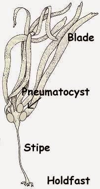
  • Made up of holdfast (base used to attach to rock or bottom), pneumatocyst (air bladder to aid buoyancy) and stipe (supporting stalk) and blade (leaflike structure that floats on the surface to collect sunlight for photosynthesis).
  • Cell walls made up of cellulose and polysaccharides called "alginic acid". 
  • Store food in the form of "laminarin". 
  • Consists about 1,500 species. 
  • Contains primary pigments : chlorophyll a and c, accessory pigments : xanthophyll and fucoxanthin.
  • Example : Fucus spp. and Sargassum spp.
Structure of brown algae 

Sargassum spp. 

Fucus spp.


Rhodophyta (Red algae) 
  • Mainly live in shallow waters and deep tropical seas. Few occurs in freshwater. 
  • Most common in warm-temperate and tropical climates.
  • Consists about 4,000 to 6,000 species. 
  • Lack flagella.
  • Store food in the form of "floridean starch". 
  • Cell walls are made up of cellulose and polysaccharides such as agar and carrageenin. 
  • Contains primary pigments : chlorophyll a and d, accessory pigments : carotenoids, xanthophylls, phycobilins
  • Example : Porolithin sp., Kappaphycus
Kappaphycus

Porolithin sp.


Euglenaphyta (euglenoids) 
  • Occurs in freshwater. 
  • Protozoa-like algae. 
  • Flagellated. 
  • Store food in the form of "paramylon". 
  • Some are photosynthetic and some are heterotrophic
  • No cell wall. 
  • Contains primary pigments : chlorophyll a and b, accessory pigments : carotenoids and xanthophylls
Structure of euglenoids

Examples of euglenoids


Chrysophyta (golden algae)
  • Occur in both marine and freshwater. 
  • Flagellated. 
  • Cell walls are made up cellulose and pectin (which often filled with silica).  
  • Store food in the form of "leucosin" and also in oil droplets. 
  • Contains primary pigments : chlorophyll a and c, accessory pigments : carotenoids, fucoxanthin, xanthopylls
A colony of golden algae. 


Dinoflagellates (Phylum Pyrrophyta) 
  • Occurs in both marine and freshwater. 
  • Some are photosynthetic, some are heterotrophic. 
  • Have two flagella. (whip, turn and maneuver in water).
  • Store food in the form of starch. 
  • Some species of dinoflagellates emit blue light when disturbed, called bioluminescence
  • Some species of dinoflagellates are toxic and can cause red tides and shellfish poisoning.
  • Contains primary pigments : chlorophyll a and c, accessory pigment : carotenoids
  • Example : Noctiluca scintillans, Pfiesteria piscicidia
Noctiluca scintillans

Pfiesteria piscicidia (SEM)
Bioluminescent algae 

Red tides (a type of harmful algal bloom)


Bacillariophyta (diatom)
  • Occurs in freshwater, salt water, moist soil and moist surface of plants. 
  • Often regarded as the most beautiful algae. 
  • cell walls are made up of glass that is very finely etched with a species-specific pattern of dots and lines. 
  • Store food in the form of chrysolaminarin (oily carbohydrate). 
  • Contains primary pigments : chlorophyll a and c, accessory pigments : carotenoids, fucoxanthin. 


USES OF ALGAE 

Algae are used in food, animal feed, cosmetics, pharmaceuticals, and biofuels. They can also be used for carbon sequestration and bioremediation of waste and waste water. (this is what I found when I Google for the uses of algae). 

While... These are the points when we discussed in class that day. Algae are used as food (seaweed), fuel, collagen (cosmetics), fertilisers, polisher, filtration and dye for textiles (clothes). 

#FUNFACT 

> Why protist is not classified as "Plantae"? 
Because they don't have vascular system like in plants. 

> Why protist is not classified as "Animal"?
Because they are autotroph (photosynthetic).


Soooo, like I said before, by the end of the blog. I should have learned something about algae. And... I did learnt something (about algae) while finishing this journal! ฅʕ•̫͡•ʔฅ 

P/S Somehow this update is more about me learning about algae instead of what I did in class that day. I'm so sorry. x'D 

Until next class. Hehe.

And happy holiday~~~ 

Adieu!

Friday, October 10, 2014

Friday, 10th October 2014, 9th Microbiology Class "Exam"

Today I arrived quite late to the exam venue. At... If I'm not mistaken, 8.35am. Fortunately, when we arrived the others were also going into the room where our very first Microbiology examination will be held. Yes. We had our examination today!! Haha. The examination is done in 1 hour and 30 minutes. 

And... I got correct for the question on what is the function for pili. Actually, we have to match the organelle with it's function. And there are some that was quite confusing and I'm not so sure about their function. 

(By the way, the answer for what's the function of pili for today's exam is ---> infecting other organisms

Afterwards, Dr Wan said that after she observed how we... answered our Microbiology paper today. She got quite worried, i guess? Haha. And that when she asked us why didn't we remember what's the function of pili. Some of us told her that they don't remember but Dr Wan said that that's not the case. The real reason on why we couldn't remember is... We are not supposed to memorise all the organelle and it's function but it is because we do not understand what we learn during class. 

So, for today's lesson which is on Topic 5 "Protozoa", she assigned us a group work. Discuss with our groupmates on what is the best way to remember or study this topic. Such as by doing mind map, watching videos or search for additional informations on the net. After deciding on the best method, share it with the class by presenting today's topic with the method that we have chosen. 

It was veryyy interestin to see how each how each group present it with their own preferred way. Hmm, Farah Alia's group and Megat's group use the mind map method by constructing a mind map online.

Chew's group chose to watch videos in order to understand more since we can visualise it and also search for words or terms that we don't understand like... "African sleeping sickness" which is a disease that is caused by flies (at least that's what Mawardah said hehe. I'll look up for it, after writing this blog. hehe). 

Ee Lyn's group gave us few questions, and we have to fill in the blanks. Which really helps! :D 

Mashi's group chose the flash cards method, they agreed that it is easier to read notes that way especially when we are having quizzes. 

And lastly, Choy's group where they presented the "Protozoa" topic by using the capital of the characteristics, like... for example PIPA which stands for Phagocytosis, Ingestion, Pinocytosis, and Absorbtion. (See? Even I remembered it up until now, haha xD) and they even prepared some "short songs" but the lyrics is based on the characteristics for the major group of protozoa. They even made us sing along with them. We were forced to, under Choy's command. Hahaa

And as for my group, we also use the mind map method but we sort the... "biography of protozoa" (Can I even say it like that? xD) by dividing the informations into four main sections which is characteristics, movement, habitat and example. And we also use the icon method. For example, for the point "housed in shell". We presented it by putting a house drawing followed by "in" and a shell drawing. We think that it is more fun that way, suitable for us "youngins". Hehe. 

But we didn't get the chance to present our presentation today because we ran out of time, the class was dragged until 12.30pm when our class was supposed to end at 12.00pm. Hee. And it was our fault anyway; very poor time management. The preparation for the presentation should only be done in 10 minutes only (that's what Dr Wan told us before we started preparing) but apparently we took more that 10 minutes.. Hee. 

Until then, 

Adieu!  

Thursday, October 9, 2014

Tuesday, 7th October 2014, 8th Microbiology Class "I don't remember what I'm feeling that day, but I'm sure it's a positive feeling"

 Class started a bit late. 15 minutes late if I am not mistaken. Dr Wan was late by 15 minutes. Hehe. It's okay though, not that I mind x'D In addition to it, Dr Wan was late. Yes. But she arrived late while carrying a plastic bag. That sprang to mind at that moment that last friday, on our previous class before we go back for Eid al-Adha, she promised to bring 'Rendang' for us. I didn't know she actually meant it. Hehehe. But she told us afterwards that she was late because she was cooking "rendang" for us but at the last minute, she remembered that some of our class members are allergic to chicken, so she cooked fried rice for the said students. That's so... Sweet and thoughtful of Dr Wan x'D
And classes soon after, on the last section of Topic 3 which covered on Inclusions.
Since today's lesson was a bit short, I shall do the "What I learned today" or "New Things That I Learned Today". Hehe.

New Things That I Learned Today

  • Although it is well known that prokaryotes have metachromatic granules, Dr Wan told us that eukaryotes also have metachromatic granules, which is used as food storage.


  • Other than as an energy reserve and having for nutrient, sulphur granules is also used to form spore.

  • (This is not something that I just knew, but I wanted to put this in the journal too, because I am reminded about it again x'D so it's important too)

  • The other name or term used for "carbon fixation" is Kelvin cycle or Kelvin-Benson cycle (Thanks to Dr Wan for reminding us!!)


  • Dr Wan asked us what is "microbial nutrition" (what they eat to survive?)

  1. Source of carbon
  2. Source of energy


  • For microbial that uses carbon dioxide and light, it is called "phototrophic microbe" or "photosynthetic".
  • While other microbial that use other kinds of chemical (other than dioxide and light), it is called "heterotrophs".

  • The function of magnetosomes is to passively align along the Earth's magnetic field lines to direct themselves to the side where there is oxygen.

  • Microbes that requires oxygen to survive is called "microaerophile".

  • Magnetosomes can be in chain form, diamond or bullet. ONLY ONE or TWO chain!
  • Other function of magnetosome is to protect the cell against hydrogen peroxide accumulation.

  • Why do the cell want to prevent from hydrogen peroxide? (Chew, one of my classmate asked that to Dr Wan, and Dr Wan asked us back. Haha. But of course we remained silent. Hehe.)

  • Some fact about hydrogen peroxide that was mentioned by Dr Wan that day.

  1. Hydrogen peroxide contained in hair dye.
  2. Aerobic organism are able to neutralise the toxicity of H
    2
    O
    2
     because they have the enzyme that help degrade H
    2
    O
    2
    .
  3. On the other hand, bacteria have magnetosome to protect themselves. While bacteria that don't have magnetosomes, have enzyme peroxidase instead.


  • When bacteria face a harsh environmental condition, it will trigger the formation of endospores.  (vegetative cell -----> endospore)
  • When bacteria face a favourable condition, it will germinate again into vegetative cell.

  • When endospore is stained with malachite green (the only stain that can detect endospore), positive result will stained green, while negative result will stained red or pink or white.

  • Desiccation = extreme dryness

  • Bacteria that can live in extreme dryness is called "xerophilic".

  • "Dipicolinic acid" is an acid that prevent from desiccation in bacteria.

  • Chew asked Dr Wan if endospore can be destroyed? Dr Wan said that certain radiation can kill it, but only radiation, no chemical can destroy it yet.


That's all for today. This is all what I can tell for today's (Monday class) journal. This is solely what Dr Wan said during class. Haha. Maybe there are some of them I have missed or didn't jot it down. Or maybe I have heard it wrongly... I hope not O_O And I didn't do any further Google-ing because the internet connection is very poor nowadays, here,at college. Hmm I don't think I can even consider it as "poor" but I couldn't even connect to the internet at all x'D We're having a bit of a problem here with the internet connection, I guess. So, there's not much information that I can share.. 

In between class, we stopped for a break. We ate the rendang and fried rice that Dr Wan brought!! It was delicious!! :D :D While on break, she asked us to "submit" what microbe that we have decided to adopt :D So... after searching and also discussing with my older sister (since she studied Medicine before, she studied Microbiology too) and she suggested some microbes but at last I picked Shigella dysenteriae after further research about some of the microbes.

I even considered diatom and volvox, like I said before (wayyy earlier, in my first or second post, maybe? x'D) but Shigella dysentriae won my heart. Keke.

And Dr Wan also introduced us to a new website, "Socrates" for us to answer some questions. It's a "pretty" website!  It's a good method to... attract our interest to answer the questions. Hehe. I'm telling this from my point of view as a student! :D
Oh and also! We can check daily news or updates about microbes on this website 

www.sciencedaily.com

Adieu!

:D

Sunday, October 5, 2014

Friday, 3rd October 2014, 7th Microbiology Class "Eid al-Adha"

Everyone was in the mood of going back home today, with their bags (please notice the (s) on the bag hahaha indicating they are carrying more bags than they usually do to class) I wish you Happy Eid al-Adha in advance :) I hope all of you arrived home safely. 

Today's lesson continues from where we last stop last week. Still under the topic of external structures of prokaryotes before proceeding on some of "The Internal structures of Prokaryotes". Despite the "Raya" mood, everybody was cooperating with the lesson as usual. Hehe. And we even had some foods mentioned during class! It's not even related to Raya actually. It's all because we came across the diagrams of peptidoglycan that looks a lot like sausages, Gram negative and Gram positive cell walls that look like spaghetti with meatballs and cytoplasm, due to it's jellylike structure, it reminds everyone of jelly or in malay we called it 'dadih'. Hahahaha. I see all of you are hungry, eh? x'D

"Spaghetti and meatballs" Gram negative and Gram positive cell walls

"Sausages" peptidoglycan 
Today Dr Wan kind of focusing more on the Gram positive and Gram negative cell walls during the end of the lesson. Thus, I shall conclude about these cell walls first.

The two cell walls are different are different by the structure itself. Like the one that I have posted above. 


GRAM POSITIVE CELL WALL 
  1. 1- Have many layers of peptidoglycan
  2. 2- Contains teichoic acid and lipoteichoic acid (which is teichoic acid that attached to plasma membrane) 
  3. 3- 90% of the cell wall is peptidoglycan. 
  4. 4- Do not contain outer membrane

FUN FACT #1

Teichoic acid
  • There are two types : wall teichoic acid (WTA) that attached to peptidoglycan and lipoteichoic acid (LTA) that attached to plasma membrane.
  • Resistance towards environmetal stresses, low osmolarity, antimicrobial peptides, antimicrobial fatty acids, cationic antibiotics and lytic enzymes produced by the host. 
  • Act as receptor for phage particles. 
  • Can bind to cationic groups thus providing a reservoir of ions close to the surface of bacteria that may ease the activity of different enzymes. 
  • Involved in cell division and cell morphogenesis (a process that concerned with the shapes of tissues, organs and entire organisms and the positions of the various specialized cell types).
  • WTA act as the spatial and temporal regulator for peptidoglycan metabolism, controlling the level of cross-linking. The highly cross-linked peptidoglycan showed a higher resistance to enzymatic degradation by lysozyme. 
  • Thus, the high resistance helped the bacteria when interacting with host that produce lysozyme as they can defense themselves against bacterial infections. 
  • And also, the cell wall does not degrade easily! 
  • Hence, that's why most of Gram positive bacteria are not pathogenic; harmless.
GRAM NEGATIVE CELL WALL 
  1. Very few layer of peptidoglycan
  2. Have outer membrane that consists of lipoproteins, lipopolysaccharides (LPS), phospholipids and porins
  3. 10% of the cell wall is peptidoglycan.
Aside from Gram positive and Gram negative cell wall, there are also atypical cell walls. Which 'atypical' itself means 'unusual'. 

ATYPICAL CELL WALL (Unusual cell wall) 
  1. Mycoplasma : Have no cell wall. Instead, they have sterols. (which has the same composition with cholesterol). 
  2. Chlamydiaceae : The cell wall contains LPS.
  3.  Archaea : Lack of peptidoglycans in their cell walls but have pseudopeptidoglycans. 
THE DIFFERENCE BETWEEN CELL WALL OF AN ARCHAEA AND BACTERIA

As I had mentioned in the previous entry. The difference between archaea and bacteria is the components of their cell wall where peptidoglycan is present in bacteria but not in archaea.

Instead, archaea have pseudopeptidoglycan which is a substance that is quite similar to peptidoglycan.

The difference between peptidoglycan and pseudopeptidoglycan is that in peptidoglycan, the disaccharide molecule is made up of monosaccharides called N-acetylglucosamine (NAG) and N-acetylmuramic acid (NAM). The alternating NAM and NAG molecules are linked by β(1,4) glycosidic bonds.

On the other hand, pseudopeptidoglycan in archaea are made up of N-acetylglucosamine (NAG) which is the same as in peptidoglycan, but instead of NAM, it has N-acetyltalosamimuronic acid (NAT).  The alternating NAM and NAT are linked by β(1,3) glysodic bonds.

Next, we proceeded on the “Internal Structures of Prokaryotes”. During this “session”, the class participated more when discussing/learning this topic, there were more response compared to when Dr Wan was teaching us about the “External Structures of Prokaryotes”. It’s obvious that this is all because of our previous assignment Dr Wan assigned us last week -->> Make a mind map for the topic of internal structures of prokaryotes.

The internal structures of prokaryotes consists of
  • Plasma membrane
  • Cytoplasm
  • The nuclear area (Nucleoid and plasmid)
  • Ribosomes
  • Inclusions
  • Endospore

FUN FACT #2

Prokaryotes do carry on respiration. There are two types of respiration which are aerobic respiration and anaerobic respiration.  The plasma membrane is the one who is responsible to carry on the respiration, though. 

PLASMA MEMBRANE

There are two movements of materials across the membrane namely the active process and passive process.

PASSIVE PROCESS

·    The process where molecules move from a region of higher concentration to a region of lower concentration, down the concentration gradient, WITHOUT the aid of energy (ATP).
·   
T   There are three types of passive process which are simple diffusion (The movement of molecules from high to low concentration until equilibrium), facilitated diffusion (the movement of molecules from high to low concentration, WITH the aid of carrier protein), and osmosis (the movement of water from high concentration of water to low concentration of water).

ACTIVE PROCESS

·    The process where molecules move from a region of lower concentration to a region of higher concentration, against the concentration gradient, WITH the aid of energy (ATP).
·   
     There are two types of active process which are active transport (molecules are moved by transporter protein from low to high concentration, with the aid of energy) and group translocation (where the transport protein change shape when transporting the molecules, with the aid of energy).

CYTOPLASM

This is where we were all reminded to “dadih” due to the jelly-like structure of the cytosol. Haha!

The major substances of cytoplasm are DNA, ribosomes and inclusions.

NUCLEAR AREA

Consists of the nucleoid and plasmid.

NUCLEOID

·    Nucleoid is a single, long circular molecule of double stranded DNA (in prokaryotes, we called it “bacterial chromosome”).

PLASMID

  •      Small, circular, double-stranded DNA.
  •      An extrachromosomal genetic elements.
  •      There are five types of plasmid with their own specific functions which are virulence plasmid, Col plasmid, fertility plasmid, degradative plasmid and resistance plasmid.
The functions of each type of plasmid

  •     They are transferable from one bacteria to another bacteria. (same species only!!)
Functions of the plasmid :

  1.     To carry antibiotic resistant genes and spread them. Thus, many diseases can be prevented or treated.
  2.    To carry genes that involved in metabolic activities.
  3.     Capable of producing antibacterial proteins. 
And there are also other functions of plasmid. 

RIBOSOMES

In my opinion, ribosome is an organelle that is very well known to everyone.  For  “everyone” that previously studied Biology/Science in secondary school, that is. Hehe.

The function of ribosome is as the site of protein synthesis. And they are one of the major components of cytoplasm, as I had mentioned earlier. In eukaryotes, some ribosome are membrane bound (where they are attached to the rough endoplasmic reticulum (RER) and some are boundless, and floating around in the cytosol). Since there are no RER in prokaryotes, the ribosomes are pretty much boundless.

There are 70S (S = Svedberg unit) ribosomes which consists of two subunits, small subunits = 30S and large subunits = 50S).

If you’re wondering why 30S and 50S doesn’t make up a total of 80S but instead of the stated 70S. I did wonder about that too, but sadly I didn’t do any initiatives to look up for it. Hee. I’ll be more productive and curious after this. I will!! And. Ah. Actually Dr Wan asked us during today’s class. “Anybody searched for why it 70S and not 80S?” but nobody answered. Hehe. 

Dr Wan said that it is because during centrifugation, there are some ribosomes that are lost, that’s why the number of ribosomes are not enough (80S).

In conclusion, cells that have a high rates of protein  synthesis, obviously have a large number of ribosomes inside them. Thus, several antibiotics will get into action in order to inhibit protein synthesis of ribosomes.

Some examples of the antibiotics are :

  •      Aminoglycoside antibiotic (streptomycin, gentamicin, azithromycin, telithromycin etc) 
  •      Macrolide antibiotics (Erythromycin, clarithromycin etc….)
FUN FACT #3

Macrolides are primarily bacteriostatic, that is by binding to 50S subunits of the ribosome, they inhibit bacterial protein synthesis. 

"Bacteriostatic" = Capable of inhibiting the growth or reproduction of bacteria. 

And today’s class ended!! Three days of holiday, is quite long…  I mean, Dr Wan said that it is long.. I… have no comment on that. Hahaha. What I do know is, I can’t wait to go home! Salam Eid al-Adha everyone! 

P/S  Although today’s lesson went well, and I understand most of it but there are some that I don’t understand, especially on the Gram positive and Gram negative part. Since there are Gram positive and Gram negative cell walls for both bacteria and archaea. It is quite confusing at first, i didn’t know that there are also Gram positive and Gram negative archaea too!! I’m surprised when I found out that in the lecture notes. Haha.. I will read more on that, read carefully. Real careful. Hmm but come to think of it, it’s not that I don’t understand. I’m confused. Hehe...